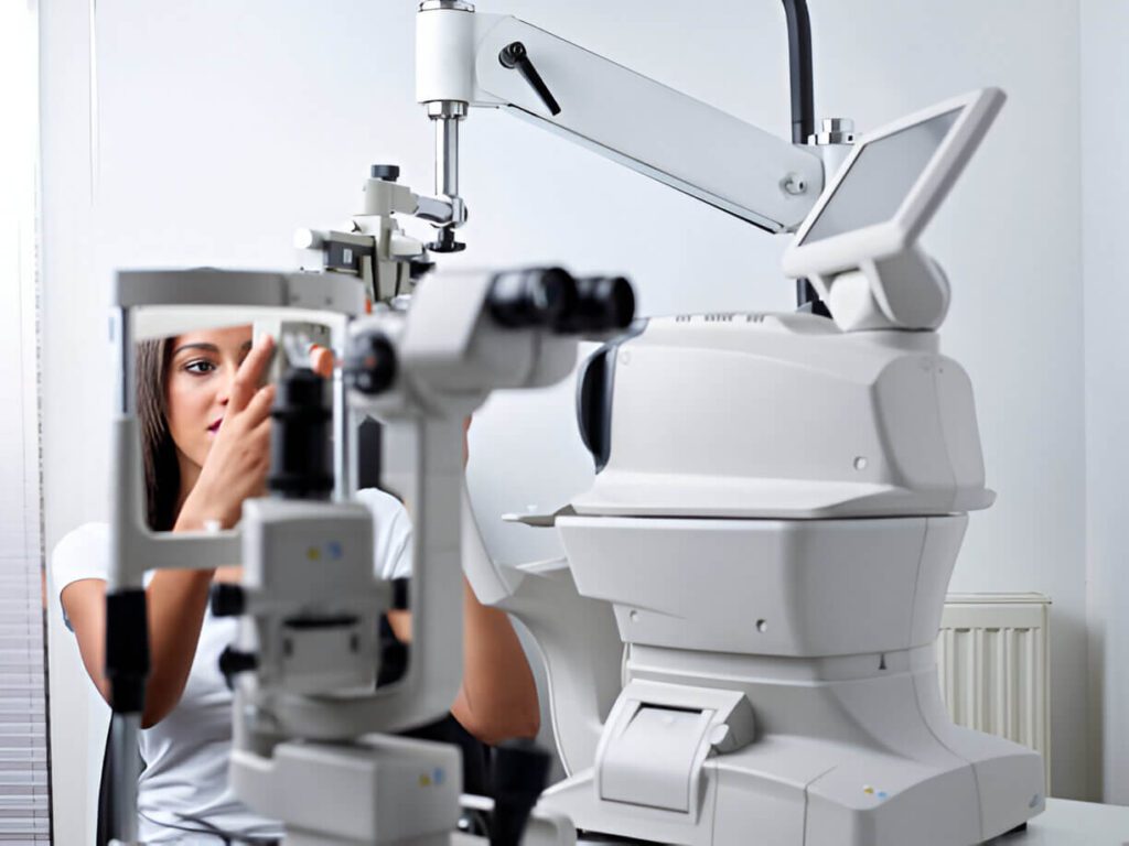
Eye surgery is delicate and needs a high level of accuracy. Even a small mistake can affect a person’s vision. That’s why eye surgeons need special tools. One of the most important tools is the ophthalmic operating microscope.
This microscope helps surgeons see tiny parts of the eye in great detail. It improves safety and success in many eye procedures. In this article, we will explain what an ophthalmic operating microscope is, how it works, and why it is important in eye care.
1. What Is an Ophthalmic Operating Microscope?
An ophthalmic operating microscope is a type of microscope made for eye surgery. It allows the surgeon to see the eye clearly during an operation. It gives a magnified and well-lit view of the surgical area.
The microscope is used in surgeries like:
- Cataract removal
- Corneal transplant
- Glaucoma surgery
- Retinal surgery
- LASIK and other laser treatments
This tool helps doctors perform these tasks with high accuracy.

2. Key Parts of an Ophthalmic Operating Microscope
These microscopes are built with many useful features. Each part plays a role in giving a clear and stable image.
Main Parts Include:
- Eyepieces – Where the surgeon looks through to view the eye
- Objective lens – Focuses on the surgical field
- Light source – Offers bright, shadow-free lighting
- Zoom and focus controls – Helps adjust magnification and clarity
- Stand or arm – Holds the microscope steady
- Foot control – Allows hands-free operation during surgery
Some microscopes also include cameras for recording the surgery.
3. How Does It Work?
The microscope is placed above the patient’s eye. The surgeon looks through the eyepieces while working. The magnified image helps the surgeon see fine details that are not visible to the naked eye.
A built-in light source shines bright light onto the eye. This helps the surgeon work with great care and accuracy.
Modern microscopes often include motorized controls. These allow the surgeon to adjust zoom and focus without using their hands.
4. Why Is It Important in Eye Surgery?
Eye surgery involves tiny parts like the cornea, lens, retina, and optic nerve. These structures are very small and delicate.
An ophthalmic operating microscope offers many benefits:
- Better visibility of small structures
- Accurate movements by the surgeon
- Lower risk of damage to the eye
- Faster recovery for the patient
- Improved results in most surgeries
Using a microscope increases the chances of a successful surgery.
5. Common Surgeries That Use This Microscope
Ophthalmic microscopes are used in many different types of eye surgery. Here are some of the most common:
Cataract Surgery
This is the removal of a cloudy lens from the eye. The microscope helps the surgeon remove and replace the lens safely.
Retinal Surgery
Used to treat retinal detachment or bleeding inside the eye. The microscope helps the doctor work deep inside the eye.
Corneal Transplant
Involves replacing a damaged cornea. The microscope ensures the new cornea is placed properly.
Glaucoma Surgery
Used to relieve pressure inside the eye. The microscope helps in making precise cuts and avoiding harm to other parts.
LASIK and Refractive Surgery
Although lasers do most of the work, a microscope is still used to examine the eye closely before and after the procedure.
6. Types of Ophthalmic Operating Microscopes
There are different types of ophthalmic microscopes. They vary based on size, control type, and technology.
Basic Manual Microscope
- Simple and affordable
- Good for small clinics
- Manual focus and light adjustment
Motorized Microscope
- Has foot pedals for zoom and focus
- Used in advanced hospitals
- Offers better control and comfort
Microscope with Camera System
- Records surgeries for training or review
- Good for teaching hospitals and large eye centers
3D Ophthalmic Microscope
- Offers a 3D view of the eye
- Used in complex surgeries
- Reduces strain on the surgeon
7. Features to Consider Before Buying
If you are planning to buy an ophthalmic operating microscope, here are some things to look for:
- Optical quality – Clear and sharp image
- Lighting system – Bright and adjustable light
- Magnification range – Should support various levels
- Ease of use – Simple controls for zoom and focus
- Ergonomics – Should be comfortable for long surgeries
- Recording options – Helpful for documentation and teaching
- Mobility – Choose between floor-mounted or ceiling-mounted models
Make sure the microscope fits your clinic or hospital’s needs.
8. Advantages of Using an Ophthalmic Operating Microscope
Let’s summarize the main benefits:
- Improves accuracy in eye surgery
- Reduces surgery time with better control
- Helps avoid complications during the procedure
- Speeds up patient recovery
- Enhances safety for both patient and surgeon
- Supports modern eye care standards
Investing in a good surgical microscope can greatly improve the quality of eye treatments.
Conclusion
An ophthalmic operating microscope is a must-have tool for eye surgeons. It allows for precise, safe, and effective treatment of many eye conditions. Whether it’s cataract removal, retinal repair, or corneal transplant, this microscope plays a key role in modern eye care.
Choosing the right microscope depends on your practice, budget, and the types of surgeries you perform. With the right tool, you can improve patient outcomes and perform surgeries with more confidence.
If you are involved in eye care, understanding and investing in this technology can take your practice to the next level.

